
September 2023
Monthly Digital Case Study
September Slides
Anemia
2023 Medical Device Network Excellence Awards
Quiz
(PDF for print)
FBC Results
WBC 30.53* (10^3/mm3)
RBC 3.95 (10^6/mm3)
HGB 12.2 (g/dL)
HCT 35.7 (%)
MCV 90 (fL)
MCH 30.8 (pg)
MCHC 34.1 (g/dL)
PLT 226* (10^3/mm3)
Neutrophils 90%
Lymphocytes 0.0%
Monocytes 6.6%
Eosinophils 0.0%
Basophils 0.0%
Metamyelocytes 3.3%
Clinical Details
Female (68 years old)
Slide Information
Intensive care unit. Flag on the platelet result on analyzer. Lymphopenia. Partially degranulated granular lineage (+). Lymphopenia. Neutrophilia. Neutrophils rather hyposegmented (++), with presence of "band cells"/metamyelocytes. Presence of macroplatelets and some platelet aggregates.
Expert Comments: Myelodysplasia/myeloproliferative syndrome?
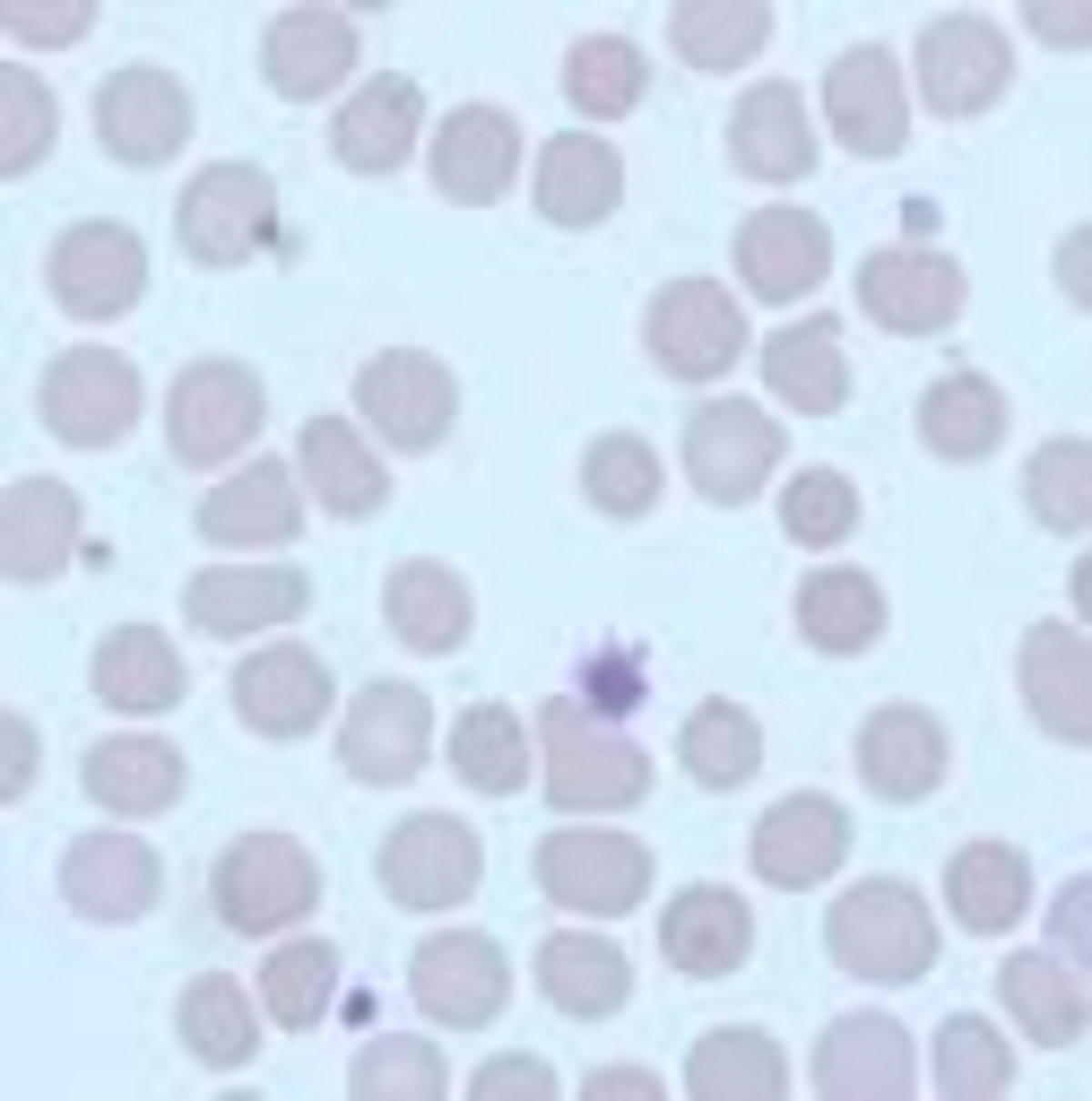
Neutrophil Band Cell
Macroplatelet
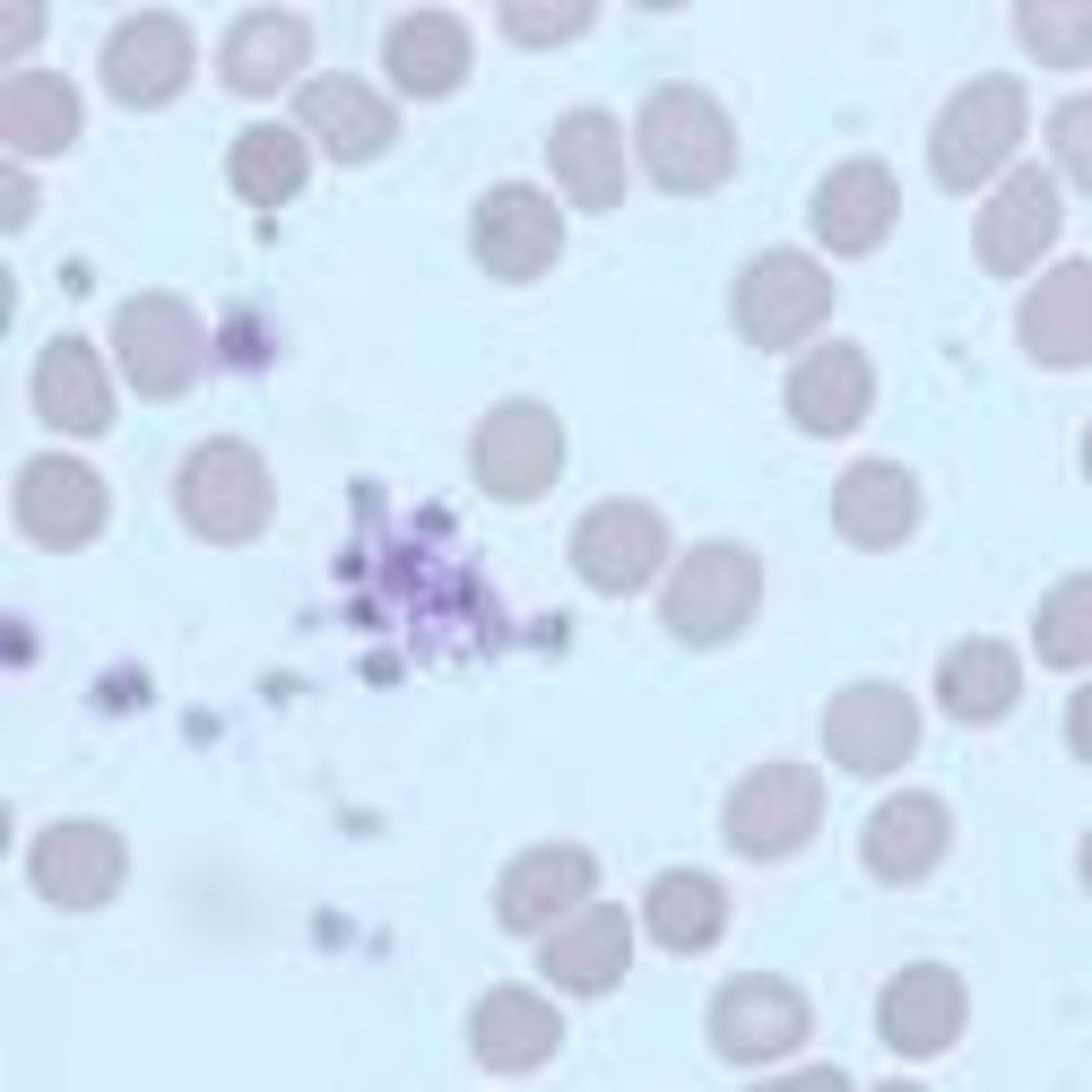
Neutrophil Band Cell
Platelet clumping
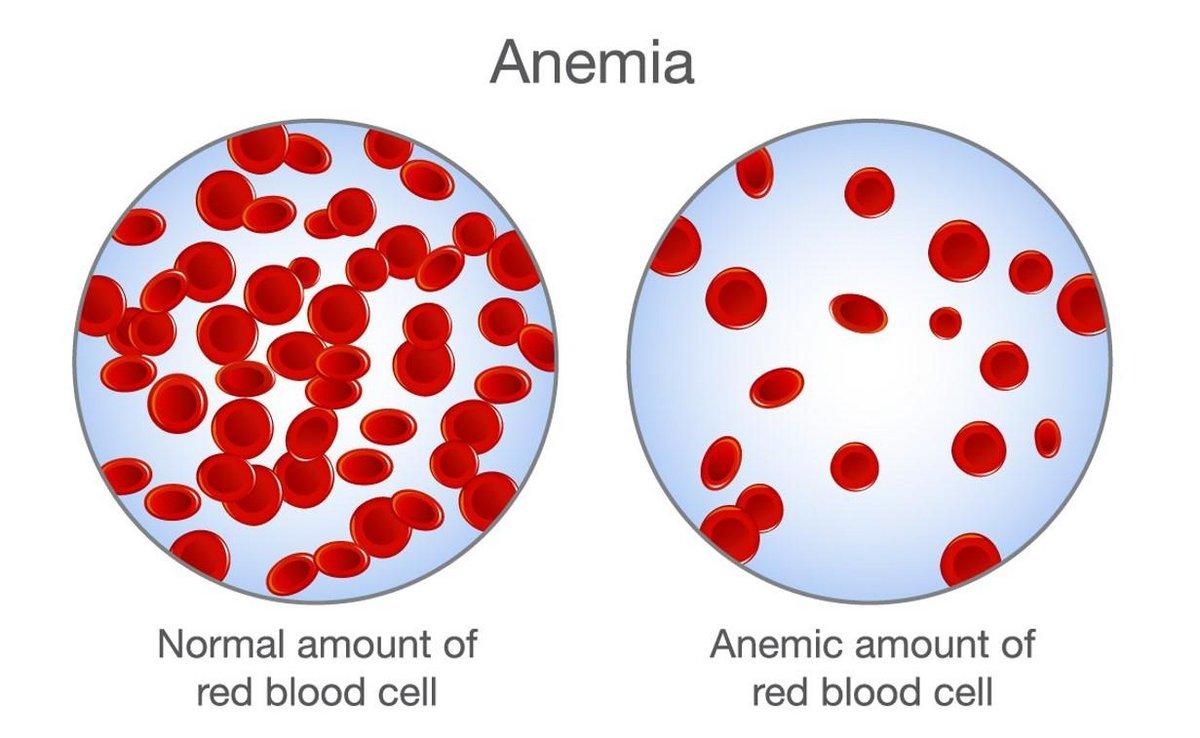
Anemia is a condition in which the blood is not carrying sufficient healthy red cells in its circulation, and therefore reduced amounts of hemoglobin carrying oxygen around the body. Anemia maybe defined as a reduced absolute number of circulating red cells, reduced hemoglobin, reduced hematocrit.
Symptoms of anemia can develop slowly, with symptoms being vague and non-specific. Symptoms that may occur first include:
When Anemia is acute, symptoms can include feeling faint and increased thirst. Symptoms of anemia depend on how rapidly the hemoglobin drops and vary depending on the underlying cause. These are usually blood loss, decreased red blood cell production, and/or increased red blood cell breakdown.
The Full Blood Count - first step to diagnosing an anemia:
In clinical workup, the MCV will be one of the first pieces of information available:
Limitations of MCV include cases where the underlying cause may be due to several factors, such as iron deficiency (a cause of microcytosis) and vitamin B12 deficiency (a cause of macrocytosis) where the net result can be normocytic cells.
In the morphological approach, anemia is classified by the size of red blood cells; this is either done automatically or on microscopic examination of a peripheral blood smear.
The causes of anemia may be classified as impaired red blood cell (RBC) production, increased RBC destruction (hemolytic anemia), blood loss and fluid overload (hypervolemia). Some of these may interplay to cause anemia. The most common cause of anemia is blood loss, but this usually does not cause any lasting symptoms, unless a relatively impaired RBC production develops, in turn, most commonly by iron deficiency.
See following newsletters for more on specific anemias.
We are proud to announce that our Yumizen hematology analyzers have won the 2023 Medical Device Network Excellence Award in the category for Environmental, Innovation, and Product Launches.
A recently updated compact instrument range, comprising the Yumizen H500 (open and closed tube sampling options) and the Yumizen H550 (autoloading) is a winner in the Innovation and the Product launch categories.
The analyzers are suited to a wide range of environments from small labs to clinics and blood banks. Read more.
Bibliography
askhematologist.com/category/anemias/
www.who.int/health-topics/anaemia
Essential Haematology, Hoffbrand & Pettit
У вас есть вопросы или пожелания? Используйте эту форму, чтобы связаться с нашими специалистами.


