X-ray Analytical Microscope Super Large Chamber Model (Micro-XRF) XGT-9000SL - HORIBA
There is a local version available of this page. Change to the local version?
United States

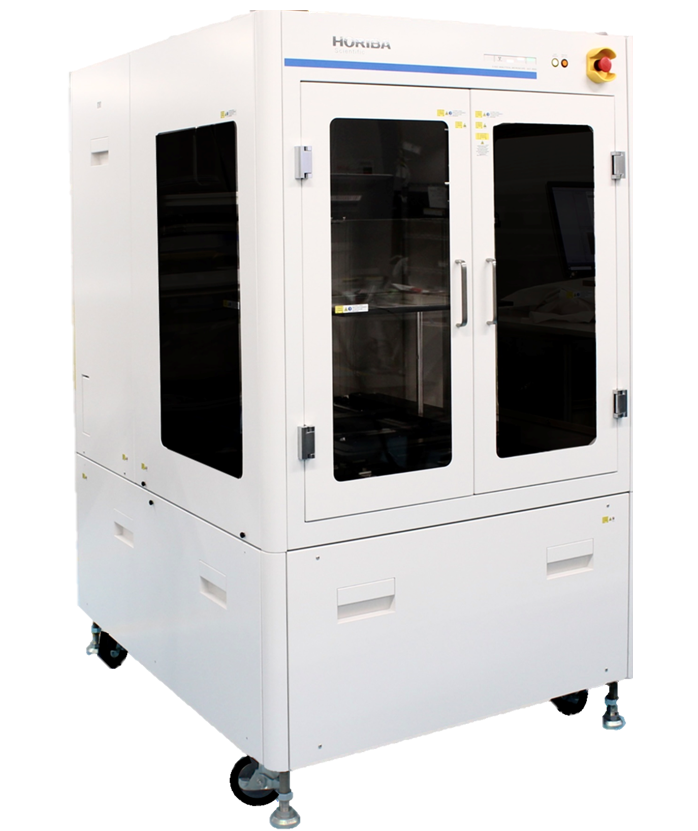
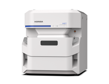
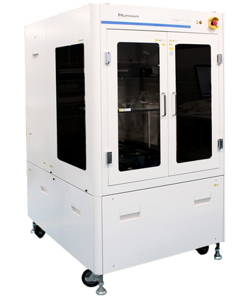
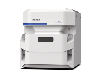
XGT-9000SL

X선 분석 현미경 초대형 챔버 모델

큰 샘플에 대한 안전한 X-선 현미경 분석 가능

  • 대용량 챔버 용량(W x D x H) : 1030 mm x 950 mm x 500 mm
  • 맵핑 영역 크기 :  350 x 350 mm2
  • 감도와 공간 분해능을 저하시키지 않는 고감도의 <15 µm 스팟 사이즈
  • 투과상 및 형광 X선 동시 검출
  • JAIMAS0101-2001/IEC1010-1을 준수하는 X선 차폐로 X선 노출로부터 사용자 보호
  • He 퍼지 모듈로 C를 비롯한 경원소 검출 가능
Segment: Scientific
Manufacturing Company: HORIBA, Ltd.

How the XGT-9000SL provides the ultimate performance in speed and flexibility

Excitation system

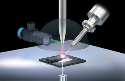
XGT-9000SL allows X-ray fluorescence analysis at the microscopic level even on a large sample by using microprobes such as <15 µm and <100 µm ultra-high intensity probes. The X-ray generator can offer high output of X-ray irradiation up to 50 kV and 1000 µA. Thus, the high performance of the excitation system provides ultimate performance and flexibility for your large sample analysis.

Optical images

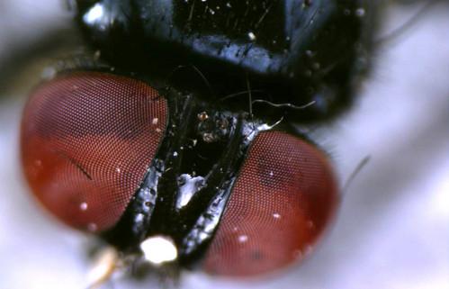
Capturing clear images is critical for micro-XRF. XGT-9000SL provides two types of cameras to grasp the image of the whole sample and its details down to the microscopic level. Multiple illumination modes help to observe brilliant images, even for reflective and transparent samples.

Sample chamber

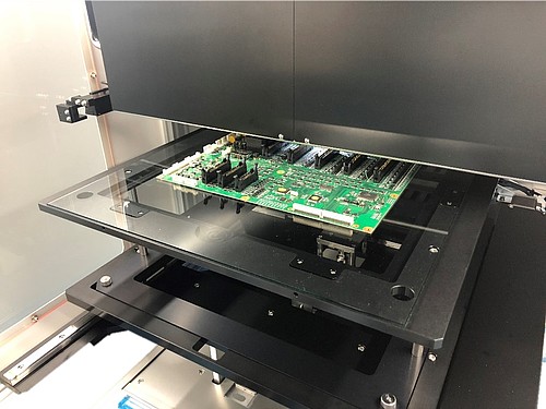
XGT-9000SL’s large chamber capacity (1030 mm x 950 mm x 500 mm) can accommodate a wide variety of samples from micro-size fragments to samples as large as electronic circuit boards and paintings. XGT-9000SL is equipped with X-ray shields complying with JAIMAS0101-2001/IEC1010-1, which protect you from X-ray exposure. XGT-9000SL can allow sample analysis without destructive sample preparation or compromising your safety.

Detectors

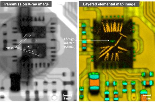
XGT-9000SL provides both a transmission X-ray detector and a fluorescent X-ray detector. The transmission X-ray detector enables users to detect internal defects and foreign matter of a sample. The fluorescent X-ray detector can detect from Americium down to carbon, with a light elements detector and He-purge module.

Software suite

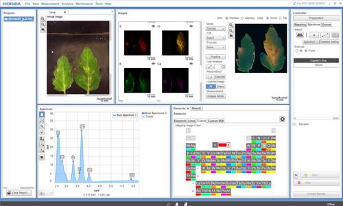
XGT-9000SL software, of course, covers all basic measurements such as single spot analysis, multiple spot analysis, line analysis, and map imaging. Apart from the standard software functions, advanced modules can be added to the software suite to provide more comprehensive user experiences. These include:

  • Multilayer FPM module for thickness measurement with/without standards
  • RoHS module for RoHS screening
  • Queue module for automated multiple measurements in unattended mode
  • Particle Finding module for particle composition/classification analysis and co-localized analysis
  • LabSpec Link module for data transfer to LabSpec 6 for multivariate analysis

scrollable

Application  Examples

Click here for more details

Basic information

InstrumentX-ray fluorescence analytical microscope
Sample typeSolids, Liquids, Particles
Detectable elementsC* – Am with optional light elements detector
F* – Am with standard detector
*He purge condition is necessary to detect down to carbon and fluorine for both detectors
Available chamber size1030(W) x 950(D) x 500(H)
Maximum sample size500(W) x 500(D) x 500(H)
Maximum mass of sample10 kg
Optical observationTwo high resolution cameras with objective lens
Optical designVertical-Coaxial X-ray and Optical observation
Sample illumination/observationTop, Bottom, Side illuminations/Bright and Dark fields
scrollable

X-ray tube

Power50 W
VoltageUp to 50 kV
CurrentUp to 1 mA
Target materialRh
scrollable

X-ray optics

Number of probesUp to 4
Primary X-ray filters for spectrum optimization5 positions
scrollable

Detectors

X-ray Fluorescence detectorSilicon Drift Detector (SDD)
Transmission detectorNaI(Tl)
scrollable

Mapping analysis

Mapping area350 mm x 350 mm
Step size4 μm
scrollable

Operating mode

Sample environmentPartial vacuum / Ambient condition / He purged condition (optional)*
*He purge condition is necessary to detect down to carbon and fluorine for both detectors.
scrollable

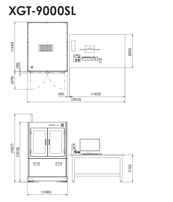
Dimensions (unit: mm)
Glass-Filled Ruby Characterization with the XGT-9000, HORIBA’s new micro-XRF
Gemstones for jewelry are treated by heating, irradiation, or filling of cracks to make them flawless. We performed imaging analyses using an XGT-9000 X-ray Analytical Microscope on a ruby provided by Mineralab. The transmission X-ray imaging revealed that the ruby included several internal defects, and the fluorescent X-ray imaging revealed that the ruby surface was treated by filling with Pb, Mg and Si which are representative of lead glass.
Non-destructive thickness and composition analysis of NiP/Au plating on Cu contacts using micro-XRF
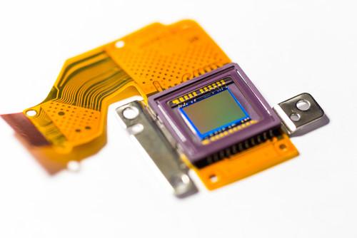
We carried out plating thickness and composition analysis on dual-layer NiP/Au plating on Cu contacts on a flexible printed circuit using a HORIBA X-ray analytical microscope. We successfully detected Au of ultra-thin layer without any sample preparation. The thickness results of Au and NiP plating were consistent with the provided values and with good repeatability.
Non-destructive failure analysis of a commercial PEMFC using a micro-XRF with a super large chamber
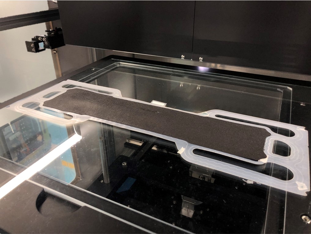
We introduce non-destructive failure analysis on a large membrane electrode assembly (MEA) from a commercial PEMFC stack using a HORIBA XGT-9000SL, a micro-XRF with an extra-large chamber capacity. Thanks to the chamber capacity, we successfully performed non-destructive elemental imaging on a defective area without cutting the sample, and we found unexpected distributions of Fe, Ni, Mn and Cr within the defective area, and that the elements are present in the MEA as a result of possible degradation of the bipolar plate.
Surface analysis on a brake rotor of automotive tire
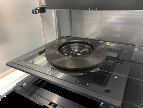
The XGT-9000SL’s large chamber capacity allows you to put whole a brake rotor and carry out spectrum analysis and elemental distribution imaging on the rotor surface, non-destructively.
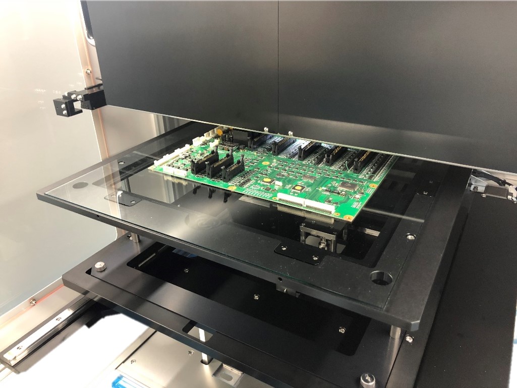
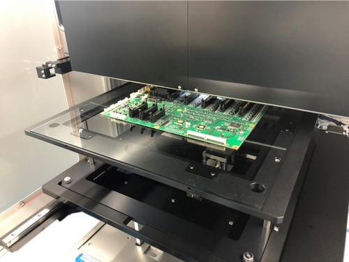
Non-destructive failure analysis on a large printed circuit board
Our XGT-9000SL allows failure analysis at a microscopic level on a large sample, non-destructively, thanks to its large chamber capacity and its microprobes.

Show More

제품 문의

HORIBA제품의 자세한 정보를 원하시면, 아래의 양식에 내용을 입력을 부탁드립니다.

* 는 필수입력항목입니다.

Related products

XGT-9000
XGT-9000

X선 분석 현미경(Micro-XRF)

XGT-9000SL
XGT-9000SL

X선 분석 현미경 초대형 챔버 모델

AP-370 series
AP-370 series

Air Pollution Monitor

AQMS
AQMS

Ambient Air Quality Monitoring System

Auto SE
Auto SE

Spectroscopic Ellipsometer for Simple Thin Film Measurement

GD-Profiler 2™
GD-Profiler 2™

Glow Discharge Optical Emission Spectrometer

LabRAM HR Evolution
LabRAM HR Evolution

Confocal Raman Microscope

LabRAM Soleil
LabRAM Soleil

Raman Microscope

LEM Series
LEM Series

Camera Endpoint Monitor based on Real Time Laser Interferometry

MESA-50
MESA-50

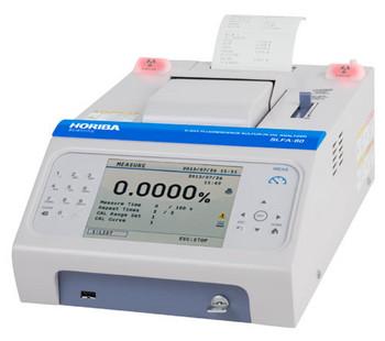
X선 형광 분석기

MESA-50K
MESA-50K

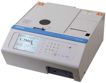
X선 형광 분석기

MEXA-2000SPCS series

배기가스 고체 입자 카운팅 시스템

OBS-ONE PM unit
OBS-ONE PM unit

차량 탑재형 배기가스 측정 장치

PX-375
PX-375

PM & 중금속 분석기

Smart SE
Smart SE

Powerful and Cost Effective Spectroscopic Ellipsometer

Ultima Expert
Ultima Expert

고해상도, 고감도, 고안정성 ICP-OES

Ultima Expert LT
Ultima Expert LT

Affordable high performance ICP-OES

UVISEL Plus
UVISEL Plus

Spectroscopic Ellipsometer from FUV to NIR: 190 to 2100 nm

XGT-9000
XGT-9000

X선 분석 현미경(Micro-XRF)

XGT-9000SL
XGT-9000SL

X선 분석 현미경 초대형 챔버 모델

XploRA™ PLUS
XploRA™ PLUS

Raman Spectrometer - Confocal Raman Microscope

Show More

MESA-50
MESA-50

X선 형광 분석기

MESA-50K
MESA-50K

X선 형광 분석기

MESA-7220V2
MESA-7220V2

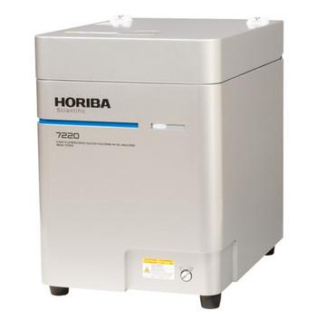
X선 형광 유중의 황/염소 분석기

PX-375
PX-375

PM & 중금속 분석기

SLFA-60
SLFA-60

X선 형광 유황 분석기

SLFA-6000
SLFA-6000

X선 형광 유황 분석기

XGT-9000
XGT-9000

X선 분석 현미경(Micro-XRF)

XGT-9000SL
XGT-9000SL

X선 분석 현미경 초대형 챔버 모델

Show More

Corporate