Clinical Case 7 - Myelodysplastic Syndrome with Excess Blasts (MDS-EB-1)
Patient demography: Male, 70’ anemia
Diagnosis: Myelodysplastic Syndrome with Excess Blasts (MDS-EB-1)
Other information: In peripheral blood - mature neutrophils (Band + Seg.) 60%, myelocytes 14%, blasts 2%, erythroblasts also present, blasts with high N/C ratio, delicate nuclear network structure and nucleoli. In addition, Pseudo-Pelger-Huët anomaly, neutrophil with hypogranularity and giant platelets were also observed, but erythroblasts showed no megaloblastoid changes. In bone marrow aspirate - giant neutrophil with hypogranularity, binucleated erythroblasts, and micromegakaryocytes. We diagnosed MDS-EB-1 based on the percentage of blasts in the peripheral blood.
Analyzer: Yumizen H2500/H1500
Case provided by: Dr. Tohru Inaba, Dept. of Infection Control and Lab. Med., Kyoto Prefectural Univ. of Medicine, Japan

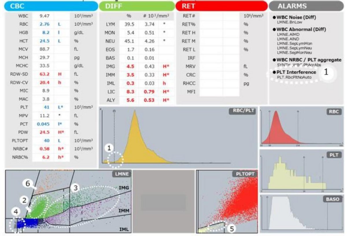
FBC results plus flagging/graphical indicators: In addition to anemia and thrombocytopenia, the RDW-SD and RDW-CV were high, suggesting abnormalities in the size and morphology of erythrocytes. Raised Large Immature Cells (LIC), especially IMG and IMM, suggesting immature myeloid cells.
➡➀ Not clearly separated, suggesting large platelets and schizocytes.
➡➁ Neutrophil population.
➡③ Reduced optical absorbance, suggesting neutrophils with hypogranularity and/ or hyposegmentation.
➡③ Populations of large immature cells are seen across the IMG and IMM regions.
➡④ Population indicating the presence of erythroblasts.
➡⑤ Area suggesting the presence of giant platelets.
