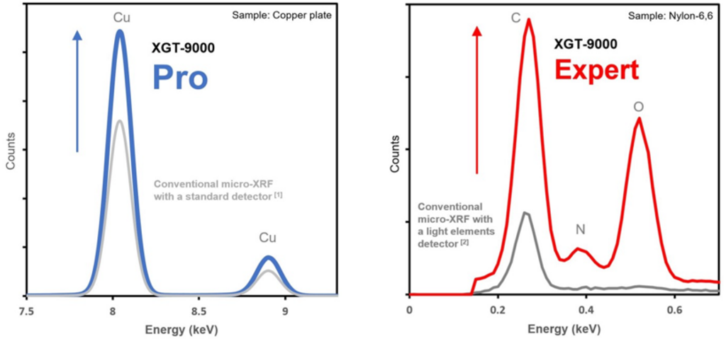
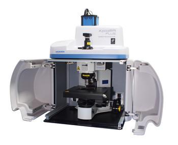
Higher sensitivity and wider detection range by new detection systems
With new advanced detection systems, the XGT-9000 Pro is able to achieve higher sensitivity than before and the XGT-9000 Expert provides you the ultimate performance in sensitivity and the widest detectable element range. Having improved sensitivity reduces measurement time and boost your work efficiency while wider detectable element range expands your application possibilities.
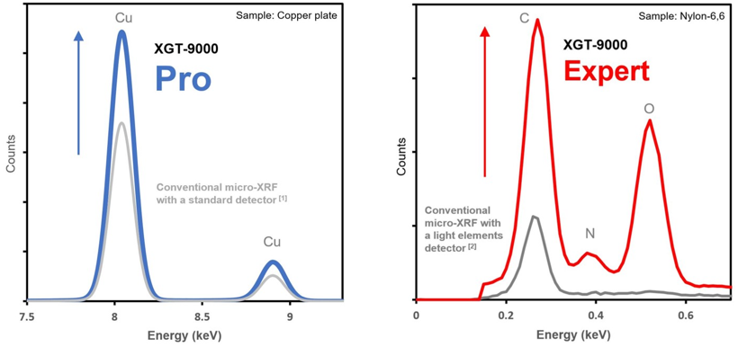
(Left) Cu intensity comparison (Right) Super light elements intensity comparison
[1][2] All the results are compared with XGT-9000 Pro/Expert vs. HORIBA conventional micro-XRF models
High quality macro and micro imaging with multiple probe selection
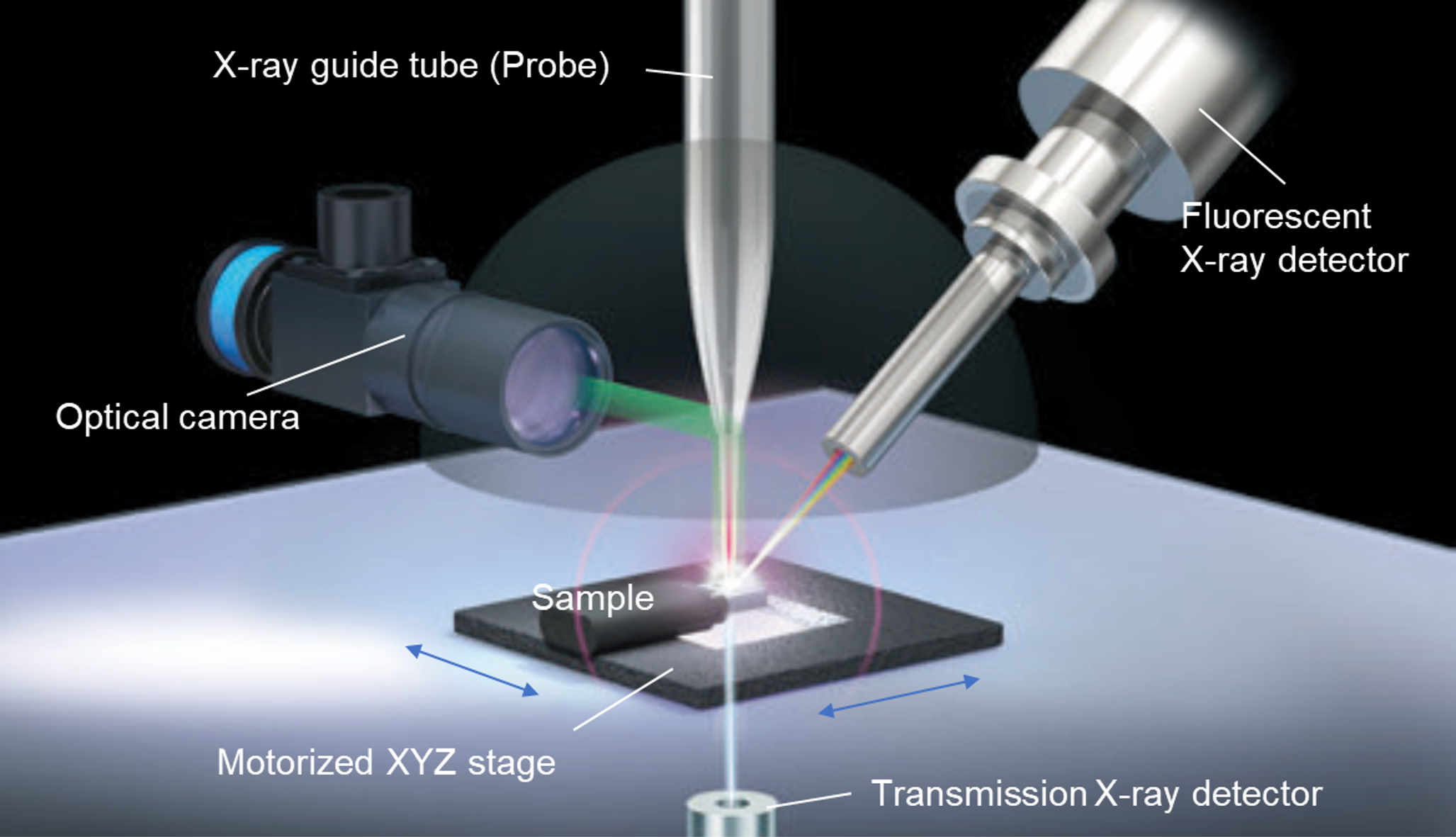
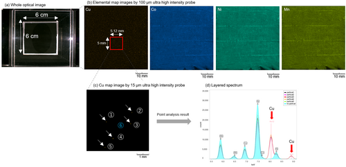
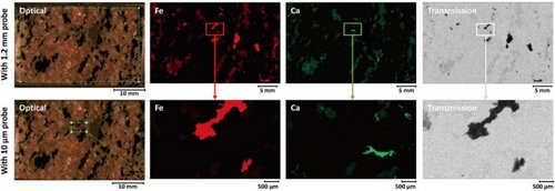
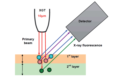
The XGT-9000 Series provides ultimate performance and flexibility with its well designed excitation system, including high output X-ray generator (up to 50 kV and 1000 µA) and a wide selection of probe spot sizes down to 10 microns and up to 1.2 mm. Multi-probes can be installed in the instrument and switchable on the software. It allows high quality fast imaging in macro and micro level without compromising the needs of spatial resolution and measurement time over large map area. Two ultra high intensity probes of 15 µm and 100 µm can be chosen.
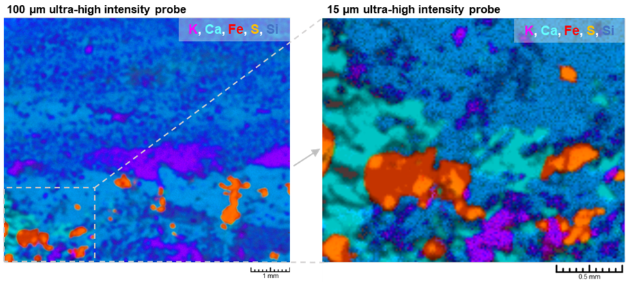
Elemental layered images on a Lapis lazuli stone using multi-probes
(Left) Fast scanning using 100 µm ultra-high intensity probe. (Right) Detailed imaging using 15 µm ultra-high intensity probe.
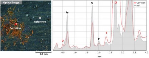
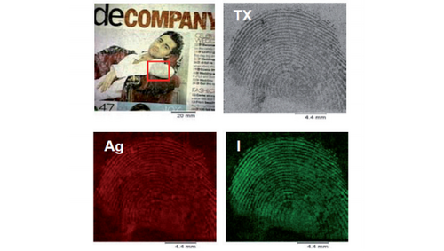
Dual types of detectors for fluorescent X-rays and transmission X-rays
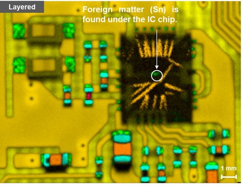
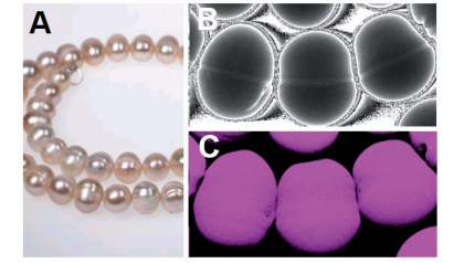
The XGT-9000 Series provides dual types of detectors. One is a fluorescent X-ray detector which tells the elemental distribution of a sample, and the other is a transmission X-ray detector which tells the internal structure of a sample. XGT-9000 can get the two types of images of the same area simultaneously. Especially it is helpful for a better understanding of electronics, gemstones including inclusions, and biological specimens.
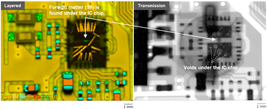
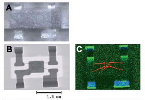
Simultaneous imaging of elemental image and transmission X-ray image of a printed circuit board
(Left) Elemental layered image revealed a foreign matter stuck under the IC chip. (Right) Transmission X-ray image revealed many voids under the IC chip.
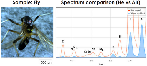
High resolution and brilliant optical images
Capturing clear images is critical for micro-XRF analysis. The XGT-9000 series provides high resolution cameras to grasp the image of the whole sample and its details down to the microscopic level. Both images can be zoomed in and out digitally to find the best position to analyze on a sample surface flexibly. In addition, thanks to the multiple illumination systems, you can get brilliant optical images of your target sample.
(Left) Whole image (Right) Detailed image
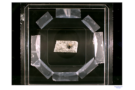
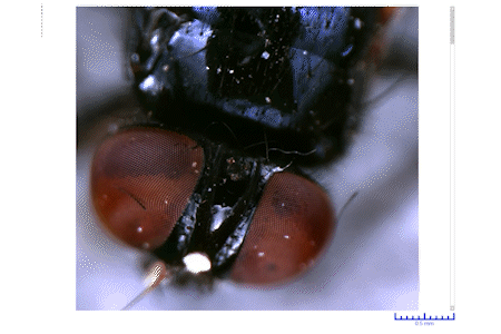
High resolution and brilliant optical image of a fly
Wide applications of the sample chamber
The XGT-9000 Series accommodates a wide variety of samples such as micro size fragments, printed circuit boards, coins, sheets, powder, liquid, and wafers. Various kinds of sample holders can be provided. In addition, up to 4 measurement environments can be selected to pull up sensitivity.
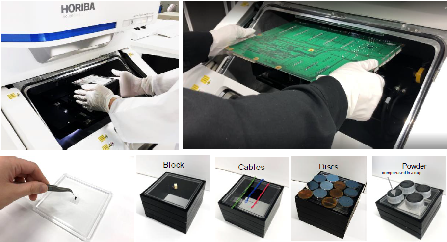
Wide applications of the sample chamber
Flexible and user-friendly software
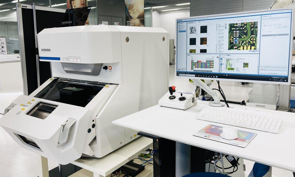
The XGT-9000 software (HORIBA X-RAY LAB) has a highly flexible and user friendly interface. It displays data tree, optical images, spectrum results, the periodic table, and map imaging results at a glance. Thanks to the flexibility, users can customize the layout on the screen. Moreover, the results can be displayed on multiple monitors. It allows you to see the results more clearly and boost your data analysis. Finally, reports can be generated, printed or exported to Excel, Word and PDF format.
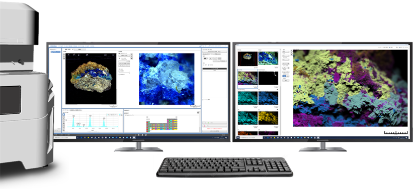
An example of software layout on dual monitors
Apart from the standard software functions, advanced modules can be added to the software suite to provide more comprehensive user experiences.
- Multilayer FPM module for thickness measurement with/without standard samples
- RoHS module for RoHS screening
- Queue module for automated multiple measurements in unattended mode
- Particle Finding module for particle analysis and co localized analysis
- LabSpec Link module for data transfer to LabSpec 6 for multivariate analysis
Publications
Application examples
Product video
Optional accessory for air-sensitive sample analysis – Transfer vessel